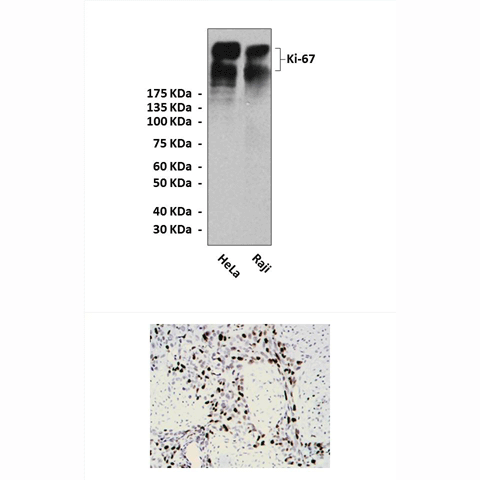
Anti-Ki-67: Mouse Ki-67 Antibody |
 |
BACKGROUND The Ki-67 antigen (also known as MKI67) is a nuclear protein that in humans is encoded by the MKI67 gene (antigen identified by monoclonal antibody Ki-67). It is strictly associated with and may be necessary for cellular proliferation.1 The expression of the human Ki-67 protein is strictly associated with cell proliferation. During interphase, the antigen can be exclusively detected within the nucleus, whereas in mitosis most of the protein is relocated to the surface of the chromosomes. The fact that the Ki-67 protein is present during all active phases of the cell cycle (G(1), S, G(2), and mitosis), but is absent from resting cells (G(0)), makes it an excellent marker for determining the so-called growth fraction of a given cell population.2 The cellular appearance and location of the Ki-67 protein throughout the cell cycle is not homogeneous. During early G1, it is found as generally weakly staining discrete foci throughout karyoplasm. It progressively condenses during late G1 in larger perinucleolar granules. During S and G2 phases, it is mainly found associated with the nucleolar region in larger foci as well as with some heterochromatin regions. When the nuclear membrane disrupts during early mitosis, Ki-67 shows an intense expression associated with the surface of condensed chromosomes in the cytoplasm. This intensity rapidly disappears in anaphase-telophase.
The Ki-67 gene is on the long arm of human chromosome 10 (10q25). Two alternative mRNA species resulting from alternative splicing encode two isoforms of the protein. The "large" Ki-67 protein isoform has a calculated molecular mass of 359 kDa, and the "small" isoform, a mass of 320 kDa. The presence or absence of the sequence encoded by exon 7 of the gene differentiates one isoform from the other. The half-life of Ki-67 protein has been estimated at around 60 to 90 minutes. Differences of expression during the cell cycle do not seem to be due to the accumulation of nondegraded proteins; rather, they seem largely to reflect variable de-novo synthesis.
Despite all this information about the nature, location, and sequence of Ki-67 protein, there is little known of its function beyond its being a protein phosphorylated via serine and threonine with a critical role in cell division. This has been concluded from the arrest of cell proliferation when Ki-67 is blocked either by microinjection of blocking antibodies or by inhibition of dephosphorylation. Furthermore it is associated with ribosomal RNA transcription. Inactivation of antigen KI-67 leads to inhibition of ribosomal RNA synthesis.
The fraction of Ki-67-positive tumor cells (the Ki-67 labeling index) is often correlated with the clinical course of cancer. The best-studied examples in this context are carcinomas of the prostate, brain and the breast. For these types of tumors, the prognostic value for survival and tumor recurrence have repeatedly been proven in uni- and multivariate analysis.3
REFERENCES
1. Urruticoechea, A. et al: J. Clin. Oncol. 23:7212-20, 2005
2. Scholzen, T. & Gerdes, J.: J Cell Physiol. 182:311-22, 2000
3. Gerdes, J.: Semin Cancer Biol. 1:199-206, 1990
2. Scholzen, T. & Gerdes, J.: J Cell Physiol. 182:311-22, 2000
3. Gerdes, J.: Semin Cancer Biol. 1:199-206, 1990
Products are for research use only. They are not intended for human, animal, or diagnostic applications.
Параметры
Cat.No.: | CP10146 |
Antigen: | Short peptide (EDLAGFKELFQTPG) from human Ki-67 protein. |
Isotype: | Mouse IgG1 |
Species & predicted species cross- reactivity ( ): | Human |
Applications & Suggested starting dilutions:* | WB 1:500 - 1:1000 IP n/d IHC 1:200 - 1:1000 ICC n/d FACS n/d |
Predicted Molecular Weight of protein: | 320/359 kDa |
Specificity/Sensitivity: | Detects endogenous Ki-67 proteins without cross-reactivity with other family members. |
Storage: | Store at -20°C, 4°C for frequent use. Avoid repeated freeze-thaw cycles. |
*Optimal working dilutions must be determined by end user.
Документы
Информация представлена исключительно в ознакомительных целях и ни при каких условиях не является публичной офертой


