Anti-S100A1: Mouse S100A1 Antibody |
 |
BACKGROUND S100 proteins are small, often acidic polypeptides of approximately 10 kDa containing an N-terminal and a C-terminal EF hand separated by a rather unstructured linker region. They were first identified in the brain and named S100 because of their solubility in 100% saturated ammonium sulfate. In humans, 19 family members are currently known with most S100 genes clustered on human chromosome 1q21 (S100A1 to S100A16). S100 proteins encoded elsewhere in the genome are S100B, S100P and S100Z. S100 proteins typically form homo- and, in some cases, also heterodimers. The dimers are thought to represent the functionally relevant entity because dimer formation appears to be required for effector protein binding. Upon binding of Ca2+, relatively large conformational changes occur, and hydrophobic surfaces are exposed in the dimer, allowing the interaction with a variety of cellular target (effector) proteins. A variety of such interaction partners have been identified, albeit in most cases still without proof of a physiological significance of the respective interaction observed in biochemical approaches. Thus, S100 proteins are considered to be signal transmitters that respond to intracellular Ca2+ rises by binding to cellular target proteins and modulating their activity.1
S100A1, a Ca2+ binding protein of the EF-hand type, is preferentially expressed in myocardial tissue and has been found to colocalize with the sarcoplasmic reticulum (SR) and the contractile filaments in cardiac tissue. S100A1 is known to modulate SR Ca2+ handling in skeletal muscle. Moreover, recent studies revealed a differential expression of S100A1 with diminished protein levels in cardiomyopathy, whereas in compensated hypertrophy S100A1 protein was found to be up-regulated. Thus, alterations in the expression of this protein might very well contribute to impaired sarcoplasmic Ca2+ handling in end-stage heart failure.2 It is suggested that S100A1 might be involved in the regulation of cardiac contractility. S100A1 improves cardiac contractile performance both by regulating SR Ca2+ handling and myofibrillar Ca2+ responsiveness.3 In addition it has been shown that the S100A1 protein is released into the extracellular space in considerable amounts during ischemic myocardial injury via an unknown mechanism. he extracellular S100A1 protein can protect cardiomyocytes from apoptosis based on Ca2+-dependent clathrin-mediated endocytotic uptake resulting in a specific activation of the pro-survival ERK1/2 signaling pathway.4 Moreover, S100A1 acts as a potent molecular chaperone and a new cochaperone protein of the Hsp70/Hsp90 multichaperone complex.
S100A1, a Ca2+ binding protein of the EF-hand type, is preferentially expressed in myocardial tissue and has been found to colocalize with the sarcoplasmic reticulum (SR) and the contractile filaments in cardiac tissue. S100A1 is known to modulate SR Ca2+ handling in skeletal muscle. Moreover, recent studies revealed a differential expression of S100A1 with diminished protein levels in cardiomyopathy, whereas in compensated hypertrophy S100A1 protein was found to be up-regulated. Thus, alterations in the expression of this protein might very well contribute to impaired sarcoplasmic Ca2+ handling in end-stage heart failure.2 It is suggested that S100A1 might be involved in the regulation of cardiac contractility. S100A1 improves cardiac contractile performance both by regulating SR Ca2+ handling and myofibrillar Ca2+ responsiveness.3 In addition it has been shown that the S100A1 protein is released into the extracellular space in considerable amounts during ischemic myocardial injury via an unknown mechanism. he extracellular S100A1 protein can protect cardiomyocytes from apoptosis based on Ca2+-dependent clathrin-mediated endocytotic uptake resulting in a specific activation of the pro-survival ERK1/2 signaling pathway.4 Moreover, S100A1 acts as a potent molecular chaperone and a new cochaperone protein of the Hsp70/Hsp90 multichaperone complex.
REFERENCES
1. Schäfer, B.W. & Heizmann, C.W.:Trends Biochem. Sci. 21:134-40, 1996
2. Remppis, A. et al: Biochim. Biophys. Acta-Mol. Cell Res. 1313:253-7, 1996
3. Most, P. et al: Proc. Natl. Acad. Sci. USA 98:13889-94, 2001
4. Most, P. et al: J. Biol. Chem. 278:48404-12, 2003
2. Remppis, A. et al: Biochim. Biophys. Acta-Mol. Cell Res. 1313:253-7, 1996
3. Most, P. et al: Proc. Natl. Acad. Sci. USA 98:13889-94, 2001
4. Most, P. et al: J. Biol. Chem. 278:48404-12, 2003
Products are for research use only. They are not intended for human, animal, or diagnostic applications.
Параметры
Cat.No.: | CP10221 |
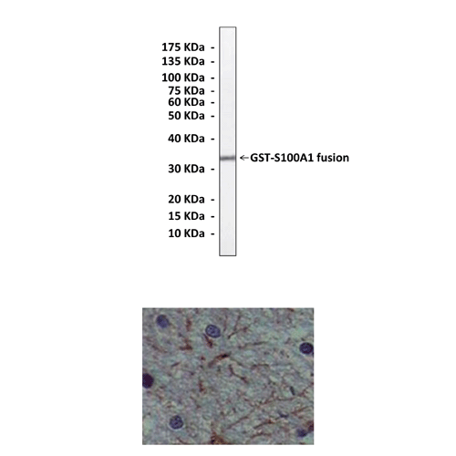
Antigen: | Purified recombinant human S100A1 proteins expressed in E. coli. |
Isotype: | Mouse IgG1 |
Species & predicted species cross- reactivity ( ): | Human |
Applications & Suggested starting dilutions:* | WB 1:1000 IP n/d IHC 1:200 ICC n/d FACS n/d |
Predicted Molecular Weight of protein: | 11 kDa |
Specificity/Sensitivity: | Detects S100A1 proteins without cross-reactivity with other family members. |
Storage: | Store at -20°C, 4°C for frequent use. Avoid repeated freeze-thaw cycles. |
*Optimal working dilutions must be determined by end user.
Документы
Информация представлена исключительно в ознакомительных целях и ни при каких условиях не является публичной офертой


