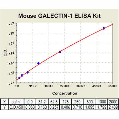
Galectin-1 ELISA Kit, Mouse |
 |
BACKGROUND Galectins are a family of animal lectins that bind beta-galactosides. Outside the cell, galectins bind to cell-surface and extracellular matrix glycans and thereby affect a variety of cellular processes. However, galectins are also detectable in the cytosol and nucleus, and may influence cellular functions such as intracellular signalling pathways through protein-protein interactions with other cytoplasmic and nuclear proteins. Current research indicates that galectins play important roles in diverse physiological and pathological processes, including immune and inflammatory responses, tumour development and progression, neural degeneration, atherosclerosis, diabetes, and wound repair.1
Galectin-1 (Gal-1) is a member of the beta-galatoside-binding lectin family and exists as a noncovalent homodimer composed of two carbohydrate recognition domains to recognize a wide range of glycoprotein or glycolipid. Gal-1 acts extracellularly by binding to cell surface receptors and extracellular matrix, such as neuropilin-1, integrins, fibronectin, laminin, and carcinoembryonic antigen. In addition, it exists intracellularly and interacts with cytoplasmic and nuclear proteins to regulate cell transformation or pre-mRNA splicing.2 Galectin-1 (Gal-1) is differentially expressed by various normal and pathological tissues and appears to be functionally polyvalent, with a wide range of biological activity. Evidence points to Gal-1 and its ligands as one of the master regulators of such immune responses as T-cell homeostasis and survival, T-cell immune disorders, inflammation and allergies as well as host–pathogen interactions. Gal-1 expression or overexpression in tumors and/or the tissue surrounding them must be considered as a sign of the malignant tumor progression that is often related to the long-range dissemination of tumoral cells (metastasis), to their dissemination into the surrounding normal tissue, and to tumor immune-escape. The possible mechanisms of how Gal-1 contributes to cancer progression and metastasis have been proposed: it regulates tumor cell growth, triggers the death of infiltrating T cells, suppresses T-cell-derived proinflammatory cytokine secretion, mediates cell-cell or cell-extracellular matrix adhesion, is involved in tumor angiogenesis, and promotes cancer cell migration Furthermore, it was shown that Gal-1 promoted tumor invasion mainly by up-regulating matrix metalloproteinase (MMP)-9 and MMP-2 and by reorganizing actin cytoskeleton. Gal-1 enhanced the activation of Cdc42, a small GTPase and member of the Rho family, thus increasing the number and length of filopodia on tumor cells.2 In addition Gal-1 in its oxidized form plays a number of important roles in the regeneration of the central nervous system after injury. The targeted overexpression (or delivery) of Gal-1 should be considered as a method of choice for the treatment of some kinds of inflammation-related diseases, neurodegenerative pathologies and muscular dystrophies. In contrast, the targeted inhibition of Gal-1 expression is what should be developed for therapeutic applications against cancer progression. Gal-1 is thus a promising molecular target for the development of new and original therapeutic tools.3
Galectin-1 (Gal-1) is a member of the beta-galatoside-binding lectin family and exists as a noncovalent homodimer composed of two carbohydrate recognition domains to recognize a wide range of glycoprotein or glycolipid. Gal-1 acts extracellularly by binding to cell surface receptors and extracellular matrix, such as neuropilin-1, integrins, fibronectin, laminin, and carcinoembryonic antigen. In addition, it exists intracellularly and interacts with cytoplasmic and nuclear proteins to regulate cell transformation or pre-mRNA splicing.2 Galectin-1 (Gal-1) is differentially expressed by various normal and pathological tissues and appears to be functionally polyvalent, with a wide range of biological activity. Evidence points to Gal-1 and its ligands as one of the master regulators of such immune responses as T-cell homeostasis and survival, T-cell immune disorders, inflammation and allergies as well as host–pathogen interactions. Gal-1 expression or overexpression in tumors and/or the tissue surrounding them must be considered as a sign of the malignant tumor progression that is often related to the long-range dissemination of tumoral cells (metastasis), to their dissemination into the surrounding normal tissue, and to tumor immune-escape. The possible mechanisms of how Gal-1 contributes to cancer progression and metastasis have been proposed: it regulates tumor cell growth, triggers the death of infiltrating T cells, suppresses T-cell-derived proinflammatory cytokine secretion, mediates cell-cell or cell-extracellular matrix adhesion, is involved in tumor angiogenesis, and promotes cancer cell migration Furthermore, it was shown that Gal-1 promoted tumor invasion mainly by up-regulating matrix metalloproteinase (MMP)-9 and MMP-2 and by reorganizing actin cytoskeleton. Gal-1 enhanced the activation of Cdc42, a small GTPase and member of the Rho family, thus increasing the number and length of filopodia on tumor cells.2 In addition Gal-1 in its oxidized form plays a number of important roles in the regeneration of the central nervous system after injury. The targeted overexpression (or delivery) of Gal-1 should be considered as a method of choice for the treatment of some kinds of inflammation-related diseases, neurodegenerative pathologies and muscular dystrophies. In contrast, the targeted inhibition of Gal-1 expression is what should be developed for therapeutic applications against cancer progression. Gal-1 is thus a promising molecular target for the development of new and original therapeutic tools.3
REFERENCES
1. Yang, R.Y. et al: Expert Rev Mol Med. 10:e17, 2008
2. Wu, M-H. et al: Mol. Cancer Res. 7:311-8, 2009
3. Camby, I. et al: Glycobiol. 16:137R-157R, 2006
2. Wu, M-H. et al: Mol. Cancer Res. 7:311-8, 2009
3. Camby, I. et al: Glycobiol. 16:137R-157R, 2006
Products are for research use only. They are not intended for human, animal, or diagnostic applications.
Параметры
Cat.No.: | CL0763 |
Target Protein Species: | Mouse |
Range: | 156pg/ml-10,000pg/ml |
Specificity: | No detectable cross-reactivity with any other cytokine. |
Storage: | Store at 4°C. Use within 6 months. |
ELISA Kits are based on standard sandwich enzyme-linked immunosorbent assay technology. Freshly prepared standards, samples, and solutions are recommended for best results.
Документы
Информация представлена исключительно в ознакомительных целях и ни при каких условиях не является публичной офертой


