SDF-1 ELISA Kit, Mouse |
 |
BACKGROUND Stromal cell–derived factor (SDF)-1/CXCL12 is a peptide chemokine initially identified in bone marrow–derived stromal cells and now recognized to be expressed in stromal tissues in multiple organs. The SDF-1 receptor, CX chemokine receptor (CXCR)4, is highly specific for SDF-1; the SDF-1/CXCR4 ligand-receptor pair is uniquely without crosstalk with other chemokines or receptors. SDF-1 and CXCR4 modulate cell migration and survival during development and tissue remodeling. A major function of the SDF-1/CXCR4 axis is chemoattraction during leukocyte trafficking and stem cell homing, in which local tissue gradients of SDF-1 attract circulating hematopoietic and tissue-committed somatic stem cells. SDF-1 is a potent chemotactic factor for T cells, monocytes, B cells, dendritic cells, mast cells, eosinophils and CD34+ haematopoietic progenitors. SDF-1 regulates homing of haematopoietic stem cells to the bone marrow, megakaryocyte transepithelial migration, platelet aggregation, and differentiation of early B cell, megakaryocytic and erythroid lineages. It also induces endothelial and neuronal migration.1 Additionally, The SDF-1/CXCR4 axis is involved in broader aspects of development, tissue repair and regeneration, and cancer. SDF-1 expressed in bone marrow inhibits the apoptosis of myeloid progenitor cells and promotes their survival. CXCR4 is expressed in human embryonic stem cells destined to become endoderm. Malignant cancer cells also express CXCR4, and their survival and migration to distant tissues is promoted by SDF-1. Experimental disruption in mice of either the SDF-1 or CXCR4 genes results in late embryonic lethality with multiple generalized developmental defects in organogenesis. SDF-1 is expressed in both endothelial and mesenchymal cells. Cultured primary endothelial cells express SDF-1, where it is required for the regulation of branching morphogenesis. However, other reports suggest that endothelial cells display SDF-1 by the transcytosis of SDF-1 produced by perivascular fibroblast-like cells. SDF-1alpha (CXCL12) may promote tumor angiogenesis directly by stimulating the formation of capillary-like structures in human vascular endothelial cells or attracting endothelial stem cells.2 Interestingly, it was also shown that SDF1/CXCR4 axis represents an autocrine system that modulates brain and neuroendocrine activity.3
It was demonstrated that SDF-1 activates Src, Akt, and Erk in the ductal cells of regenerating pancreas in NOD mice and induces migration and supports survival of duct cells, suggesting that SDF-1 may be obligatory for pancreatic regeneration from cells of ductal origin. Moreover, SDF-1 stimulates Akt activity via the coupling of phosphatidylinositol (PI)-3 kinase-γ to Gi.4
It was demonstrated that SDF-1 activates Src, Akt, and Erk in the ductal cells of regenerating pancreas in NOD mice and induces migration and supports survival of duct cells, suggesting that SDF-1 may be obligatory for pancreatic regeneration from cells of ductal origin. Moreover, SDF-1 stimulates Akt activity via the coupling of phosphatidylinositol (PI)-3 kinase-γ to Gi.4
REFERENCES
1. Jaleel, M.A. et al: Mol. Hum. Reprod. 10:901-9, 2004
2. Chu, C-Y. et al: Carcinogenesis 30:205-13, 2009
3. Barbieri, F. et al: J. Mol. Endocrinol. 38:383-9, 2007
4. Sotsios, Y. et al: J. Immunol. 163:5954-63, 1999
2. Chu, C-Y. et al: Carcinogenesis 30:205-13, 2009
3. Barbieri, F. et al: J. Mol. Endocrinol. 38:383-9, 2007
4. Sotsios, Y. et al: J. Immunol. 163:5954-63, 1999
Products are for research use only. They are not intended for human, animal, or diagnostic applications.
Параметры
Cat.No.: | CL0500 |
Target Protein Species: | Mouse |
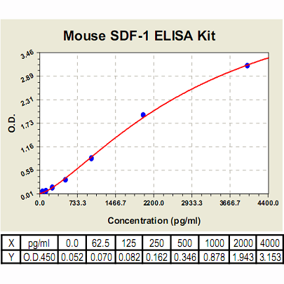
Range: | 62.5pg/ml-4000pg/ml |
Specificity: | No detectable cross-reactivity with any other cytokine. |
Storage: | Store at 4°C. Use within 6 months. |
ELISA Kits are based on standard sandwich enzyme-linked immunosorbent assay technology. Freshly prepared standards, samples, and solutions are recommended for best results.
Документы
Информация представлена исключительно в ознакомительных целях и ни при каких условиях не является публичной офертой


