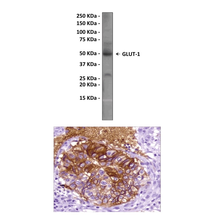
Anti-GLUT-1: Rabbit GLUT-1 Antibody |
 |
BACKGROUND Glucose transport in mammalian cells is mediated by a family of structurally related glycoproteins, the glucose transporters (GLUTs). Base on the sequence and functional similarities, GLUT family can be divided into three subfamilies, namely class I (the previously known glucose transporters GLUT1-4), class II (the previously known fructose transporter GLUT5, the GLUT7, GLUT9 and GLUT11), and class III (GLUT6, 8, 10, 12, and the myo-inositol transporter HMIT1). Functional characteristics have been reported for some of the novel GLUTs. Like GLUT1-4, they exhibit a tissue/cell-specific expression (GLUT6, leukocytes, brain; GLUT8, testis, blastocysts, brain, muscle, adipocytes; GLUT9, liver, kidney; GLUT10, liver, pancreas; GLUT11, heart, skeletal muscle). GLUT6 and GLUT8 appear to be regulated by sub-cellular redistribution, because they are targeted to intra-cellular compartments by dileucine motifs in a dynamin dependent manner. Sugar transport has been reported for GLUT6, 8, and 11; HMIT1 has been shown to be an H+/myo-inositol co-transporter. Thus, the members of the extended GLUT family exhibit surprisingly diverse substrate specificity, and the definition of sequence elements determining this substrate specificity will require a full functional characterization of all members.1
GLUT1 is ubiquitously expressed in most cells often together with other tissue-specific GLUTs, with particularly high levels in human erythrocytes and in the endothelial cells lining the blood vessels of the brain. GLUT3 is expressed primarily in neurons, and together, GLUT1 and GLUT3 allow glucose to cross the blood-brain barrier and enter neurons.2 Expression levels of GLUT1 in cell membranes are increased by reduced glucose levels and decreased by increased glucose levels. GLUT1 is responsible for the low-level of basal glucose uptake required to sustain respiration in all cells.3
Mutations in the GLUT1 gene are responsible for GLUT1 deficiency or De Vivo disease, which is a rare autosomal dominant disorder. This disease is characterized by a low cerebrospinal fluid glucose concentration (hypoglycorrhachia), a type of neuroglycopenia, which results from impaired glucose transport across the blood-brain barrier. GLUT1 is also a receptor used by the HTLV virus to gain entry into target cells.4
REFERENCES
1. Joost, H.G. & Thorens, B.: Mol Membr Biol. 18:247-56, 2001
2. Hruz, P.W. & Mueckler, M.M.: Mol. Membr. Biol. 18: 183–93, 2001
3. Knott, R.M. et al: Biochem. J. 318:313-7, 1996
4. Manel, N. et al: Cell 115:449-59, 2003
2. Hruz, P.W. & Mueckler, M.M.: Mol. Membr. Biol. 18: 183–93, 2001
3. Knott, R.M. et al: Biochem. J. 318:313-7, 1996
4. Manel, N. et al: Cell 115:449-59, 2003
Products are for research use only. They are not intended for human, animal, or diagnostic applications.
Параметры
| Cat.No.: | CG1181 |
| Antigen: | Synthetic peptide derived from the C-terminus of human GLUT-1 protein. |
| Isotype: | Rabbit IgG |
Species & predicted species cross- reactivity ( ): | Human |
Applications & Suggested starting dilutions:* | WB 1:200 IP n/d IHC 1:100-1:200 (citrate retrevial) ICC n/d FACS n/d |
Predicted Molecular Weight of protein: | 50 kDa |
| Specificity/Sensitivity: | Detects endogenous GLUT-1 proteins without cross-reactivity with other family members. |
| Storage: | Store at -20°C, 4°C for frequent use. Avoid repeated freeze-thaw cycles. |
Документы
Информация представлена исключительно в ознакомительных целях и ни при каких условиях не является публичной офертой


