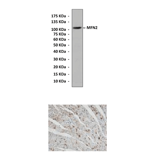
Anti-MFN2: Polyclonal Mitofusion 2 Antibody |
 |
BACKGROUND Mitofusin-2 (Mfn2) is a dynamin-like GTPase localizing to the mitochondrial outer membrane and plays an essential role in mitochondrial fusion, thus regulating mitochondrial morphology and function. Mitofusin complexes on adjacent mitochondrial membranes act as membrane tethers prior to fusion and form dimeric, antiparallel coiled-coil structures in trans. Both homo- and heterooligomeric mitofusin complexes are active. It was shown that A fraction of MFN2 present in the ER membrane assembles with MFN1 or MFN2 at the mitochondrial surface and tethers ER to mitochondria. ER-mitochondria juxtaposition is required for efficient Ca2+ uptake into mitochondria and may have important implications for cell signaling and mitochondrial movement.1
Various studies have shown that Mfn2 is a multifunctional protein that participates in cell proliferation and metabolism and that it is required for normal endoplasmic reticulum morphology. Mfn2 profoundly suppresses cell growth and proliferation in multiple tumor cell lines and rat vascular smooth muscle cells in vivo and in culture systems via inhibition of the Ras-ERK MAPK signaling pathway and down-regulation of Mfn2 contributes to various vascular proliferative disorders. In addition to its anti-proliferative effects, previous studies have shown that Mfn2 associates with Bax, a pro-apoptotic member of the Bcl-2 family, at mitochondrial scission sites during the initial stages of apoptosis, suggesting that Mfn2 might participate in mitochondrial apoptotic signaling. In contrast, emerging evidence suggests a protective effect of Mfn2 in mammalian cells. This phenomenon has been interpreted to indicate that high levels of fused mitochondria might constitute a cell defense mechanism against the accumulation of oxidative lesions.2
In relation to the metabolic role of Mfn2, alterations in activity have been reported to modify cell respiration, substrate oxidation, and oxidative phosphorylation subunit expression in cultured nonmuscle and muscle cells. Mfn2 expression in skeletal muscle is subject to regulation and conditions characterized by reduced mitochondrial activity, such as obesity or type 2 diabetes, and are associated with repressed Mfn2. In contrast, cold-exposure treatments with beta3-adrenergic agonists or exercise induce the expression of this gene in muscle. Estrogen-related receptor-alpha transcription factor is a key regulator of Mfn2 transcription and recruits peroxisome proliferator-activated receptor gamma coactivator (PGC)-1beta and PGC-1alpha. These 2 nuclear coactivators are potent, positive regulators of Mfn2 expression in muscle cells, and ablation of PGC-1beta causes Mfn2 downregulation in skeletal muscle and in the heart. Thus it is suggested that PGC-1beta is a regulator of normal expression of Mfn2 in muscle, whereas PGC-1alpha participates in the stimulation of Mfn2 expression under a variety of conditions characterized by enhanced energy expenditure.3 Furthermore, Mfn2 is involved in insulin-stimulated myogenesis. Insulin stimulates the expression of Mfn2 protein, which in turn binds to Ras and inhibits the MEK-dependent signaling pathway. At the same time, the PI3-K-dependent signaling pathway is boosted, mitochondrial respiration increases and the rate of myogenesis is accelerated.4 Additionally, mutations in MFN2 cause Charcot-Marie-Tooth (CMT) type 2A, a peripheral neuropathy characterized by axonal degeneration.5
REFERENCES
1. de Brito, O.M. & Scorrano,L.: Nature 456:605-10, 2008
2. Shen, T. et al: J. Biol. Chem. 282:23354-61, 2007
3. Zorzano, A.: App. Physiol. Nutr. Metab. 34:433-9, 2009
4. Pawlikowska, P. et al: Cell Tissue Res. 327:571-81, 2007
5. Kwan, C.C. et al: Chin. Med. J. 123:1466-9, 2010
2. Shen, T. et al: J. Biol. Chem. 282:23354-61, 2007
3. Zorzano, A.: App. Physiol. Nutr. Metab. 34:433-9, 2009
4. Pawlikowska, P. et al: Cell Tissue Res. 327:571-81, 2007
5. Kwan, C.C. et al: Chin. Med. J. 123:1466-9, 2010
Products are for research use only. They are not intended for human, animal, or diagnostic applications.
Параметры
Cat.No.: | CA1790 |
Antigen: | C-terminal sequence of human MFN2 |
Isotype: | Affinity-purified rabbit polyclonal IgG |
Species & predicted species cross- reactivity ( ): | Human, Rabbit, Rat, Mouse |
Applications & Suggested starting dilutions:* | WB 1:500 to 1:1000 IP n/d IHC (Paraffin) 1:50 to 1:200 ICC n/d FACS n/d |
Predicted Molecular Weight of protein: | 105 kDa |
Specificity/Sensitivity: | Reacts specifically with MFN2 of human, rabbit, mouse & rat origin in immunostaining and Western blot applications. |
Storage: | Store at 4° C for frequent use; at -20° C for at least one year. |
*Optimal working dilutions must be determined by end user.
Документы
Информация представлена исключительно в ознакомительных целях и ни при каких условиях не является публичной офертой


