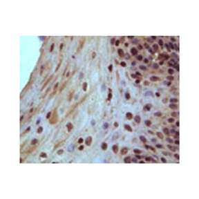
Anti-RB: Mouse Rb Antibody |
 |
BACKGROUND The Rb protein is a tumor suppressor, which plays a pivotal role in the negative control of the cell cycle and in tumor progression. It has been shown that Rb protein (pRb) is responsible for a major G1 checkpoint, blocking S-phase entry and cell growth. The retinoblastoma family includes three members, Rb/p105, p107 and Rb2/p130, collectively referred to as ‘pocket proteins’. The pRb protein represses gene transcription, required for transition from G1 to S phase, by directly binding to the transactivation domain of E2F and by binding to the promoter of these genes as a complex with E2F. pRb represses transcription also by remodeling chromatin structure through interaction with proteins such as hBRM, BRG1, HDAC1 and SUV39H1, which are involved in nucleosome remodeling, histone acetylation/deacetylation and methylation, respectively. Loss of pRb functions may induce cell cycle deregulation and so lead to a malignant phenotype. Gene inactivation of pRB through chromosomal mutations is one of the principal reasons for retinoblastoma tumor development. Functional inactivation of pRb by viral oncoprotein binding is also shown in many neoplasias such as cervical cancer, mesothelioma and AIDS-related Burkitt's lymphoma.1
RB is phosphorylated in a cell cycle-dependent manner by cyclin-dependent protein kinases (cdk’s). In quiescent and early G1 cells, RB exists in a predominantly unphosphorylated state. As cells progress toward S phase, RB becomes phosphorylated. Initial phosphorylation of RB ismost likely catalyzed by cdk4-cyclin D or cdk6-cyclin D complexes; thereafter, RB phosphorylation is maintained by other cdk-cyclins. However, it was shown that activated ERK1/2 is also responsible for mitogen induced rapid Rb phosphorylation specifically at Ser780 and Ser795. This process occurs independent of cyclin/cdk pathway.2 Many other kinases may be also involved in Rb phosphorylation, such as PKC etc.3 During mitosis, RB is rapidly dephosphorylated by an anaphase-specific phosphatase. Sixteen potential sites for cdk-mediated phosphorylation (Ser/Thr-Pro Motifs, Thr5, Ser230, Ser249, Thr252, Thr356, Thr373, Ser567, Ser608, Ser612, Ser780, Ser788, Ser807, Ser811, Thr821, and Thr826) exist in RB, and 11 of these sites have been shown to be phosphorylated in vivo.4 Different cdk-cyclin complexes preferentially phosphorylate RB at distinct sites in vitro, suggesting that different cdk-cyclin complexes may exert distinct effects on RB function in vivo. Phosphorylation of RB inactivates its growth suppression activity. In general, agents which block RB phosphorylation arrest cell cycle progression in G1, while agents which stimulate RB phosphorylation promote cell cycle progression. Phosphorylation of RB is required for G1/S progression. It was shown that p16INK4, an inhibitor of cdk4-cdk6, inhibits the phosphorylation of RB and arrests only RB-positive cells in G1. The biological significance of RB phosphorylation and its impact on the ordered progression through G1/S are exemplified by the frequent mutations that affect this process in human tumors. For example, amplification of the cyclin D1 gene is observed in breast cancer and esophageal cancer cells, in which it is believed to promote RB phosphorylation. Likewise, p16INK4, which acts to prevent RB phosphorylation, is lost in a large fraction of human tumors, again leading to the deregulated phosphorylation of RB. Additionally, the distinct phosphorylation sites are responsible for regulating the different protein binding activities of RB through different mechanisms. It was shown that phosphorylation of Thr356, Thr373, Ser-780, Ser-795, Ser-807, Ser-811, and Thr-821 decreases the binding of Rb to E2F, which would permit endothelial cells to progress fromG1 to later stages of the cell cycle.3,5 On the other hand, upon calcium influx, RB1 is dephosphorylated by calcineurin, which leads to release of the repressor complex.
REFERENCES
1. Harbour, J.M. & Dean, D.C. Nature Cell Biol. 2:E65-E67, 2000
2. Guo, J. et al: J. Biol. Chem. 280:35922-8, 2005
3. Suzuma, K. Et al: Proc. Natl. Acad. Sci. USA 99:721-6, 2002
4. Knudsen, E.S. & Wang, J.Y.J.: Mol. Cell. Biol. 17:5771-83, 1997
5. Burke, J.R. et al: J. Biol. Chem. 285:16286-93, 2010
Products are for research use only. They are not intended for human, animal, or diagnostic applications.
Параметры
Cat.No.: | CP10213 |
Antigen: | Purified recombinant human Rb fragments expressed in E. coli. |
Isotype: | Mouse IgG1 |
Species & predicted species cross- reactivity ( ): | Human |
Applications & Suggested starting dilutions:* | WB 1:1000 IP n/d IHC 1:200 ICC n/d FACS n/d |
Predicted Molecular Weight of protein: | 105 kDa |
Specificity/Sensitivity: | Detects endogenous Rb proteins without cross-reactivity with other family members. |
Storage: | Store at -20°C, 4°C for frequent use. Avoid repeated freeze-thaw cycles. |
*Optimal working dilutions must be determined by end user.
Документы
Информация представлена исключительно в ознакомительных целях и ни при каких условиях не является публичной офертой


