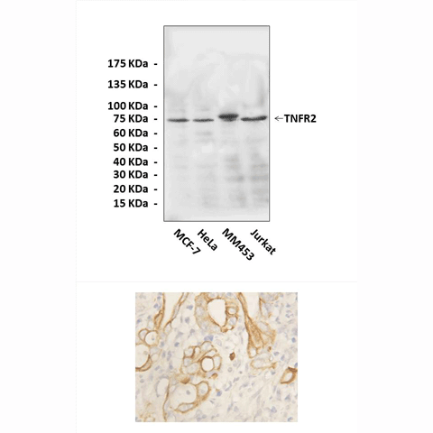
Anti-TNFR2: Rabbit TNFR2 Antibody |
 |
BACKGROUND TNF-alpha is a pleiotropic cytokine that regulates multiple biological processes. The biologic activities of TNF-alpha are mediated by two structurally related, but functionally distinct, receptors: TNFR1 (CD120alpha or p55) and TNFR2 (CD120-beta or p75). TNFR1 is the primary signaling receptor on most cell types through which the majority of inflammatory responses classically attributed to TNF occur. In contrast, TNFR2 is important in modulating TNFR1-mediated signaling, regulating autoimmune diseases, and in host defenses against specific viruses. More recently, TNFR2 has been shown to play a critical role for effective priming, proliferation, and recruitment of tumor-specific CD8 T cells.1 It was shown that TNFR2 functions as one of the earliest costimulatory members of the TNFR superfamily and TNFR2 plays a critical role in lowering the threshold for T cell activation. TNFR2 is required for sustained AKT activity and NF-kappa-B activation in response to TCR/CD28 stimulation. Moreover, TNFR2−/− T cells possess a defect in survival during the early phase of T cell activation that is correlated with a striking defect in Bcl-xL expression. These data reveal the synergistic requirement of TCR, CD28, and TNFR2 for optimal IL-2 induction and T cell survival.2 Furthermore, Many cell types coexpress TNFR1 and TNFR2 and require cooperation between these two receptors to generate TNF responses. This is exemplified in a number of cases of TNF-mediated apoptosis. The major difference between TNFR1 and TNFR2 is in their mode of signal transduction. TNFR1 belongs to a subgroup of TNFR superfamily molecules that carries a cytoplasmic death domain. Death domain TNFRs such as TNFR1 and Fas signal primarily via recruitment of death domain containing signaling molecules like TRADD, FADD, and RIP. On the other hand, non-death domain TNFRs such as TNFR2, CD30, CD40, 4-1BB, and receptor activator of NF-κB (RANK) signal by directly binding members of the TNF receptor-associated factor (TRAF) family. TRAFs bind to TNFR molecules and some other cellular proteins via their TRAF-C domain. In the case of TRAF2, all known binding sites present on TNFR molecules conform to a “major” consensus motif (P/S/A/T)X(Q/E)E, whereas the “minor” TRAF2 binding motif, PXQXXD, describes a separate class of TRAF2-binding sites found on the viral oncoprotein LMP1 and the regulatory protein I-TRAF/TANK. Based on structural data, TRAF2 is recruited to ligand-trimerized receptors via a receptor binding groove on the TRAF-C domain, whereupon it assembles into a homotrimer via the TRAF-N domain coiled-coil. Once trimerized, the amino-terminal regions of TRAFs are thought to interact with downstream effector molecules that link them to intracellular signaling pathways. These molecules include kinases such as MEKK1, NIK, and T2K and the ubiquitin-conjugating enzyme Ubc13. Unlike nearly all other non-death domain TNFR molecules, TNFR2 has only one known TRAF-binding site. Because TRAF2 but not TRAF5 or TRAF6 has been shown to bind to this site, TNFR2 appears to utilize TRAF2 for signaling.3 It has been shown that TNFR2 stimulation leads to degradation of TNF receptor associated factor-2 (TRAF2) and inhibition of TNFR1-induced activation of NFkappaB and JNK. TRAF1 inhibits TNFR2-induced proteasomal degradation of TRAF2 and relieves TNFR1-induced activation of NFkappaB from the inhibitory effect of TNFR2. Thus, TRAF1 shifts the quality of integrated TNFR1-TNFR2 signaling from apoptosis induction to proinflammatory NFkappaB signaling.4
REFERENCES
1. Watts, T.H.: Ann. Rev. Immunol. 23:23-68, 2005
2. Kim, E.Y. et al: J. Immunol. 183:6051-7, 2009
3. Grech, A.P. et al: J. Biol. Chem. 280:31572-81, 2005
4. Wicovsky A. et al: Oncogene 28:1769-81, 2009
2. Kim, E.Y. et al: J. Immunol. 183:6051-7, 2009
3. Grech, A.P. et al: J. Biol. Chem. 280:31572-81, 2005
4. Wicovsky A. et al: Oncogene 28:1769-81, 2009
Products are for research use only. They are not intended for human, animal, or diagnostic applications.
Параметры
Cat.No.: | CA1243 |
Antigen: | Short peptide from human TNFR2 sequence. |
Isotype: | Rabbit IgG |
Species & predicted species cross- reactivity ( ): | Human, Mouse, Rat |
Applications & Suggested starting dilutions:* | WB 1:1000 IP n/d IHC 1:50 - 1:200 ICC n/d FACS n/d |
Predicted Molecular Weight of protein: | 75 kDa |
Specificity/Sensitivity: | Detects endogenous levels of TNFR2 proteins without cross-reactivity with other related proteins. |
Storage: | Store at -20°C, 4°C for frequent use. Avoid repeated freeze-thaw cycles. |
*Optimal working dilutions must be determined by end user.
Документы
Информация представлена исключительно в ознакомительных целях и ни при каких условиях не является публичной офертой


