| |
BrainFectIN |
 |
BrainFectIN™ enables nucleic acids delivery into central nervous system of small animals.
Major difficulties with gene delivery in the central nervous system is the weakness of standard non-viral gene carriers and the limitations associated to the use of viral particles (time-consuming and requires additional saftey precautions). Unlike these methods, BrainFectIN™ is an original non-viral formulation that allows safe, easy and efficient nucleic acids delivery into central nervous system of small animals.
This transfection reagent allows transfection of neural cells in specific brain following stereotaxic injection, with low immunogenicity and rapid and long-term transgene expression.
BrainFectIN™ has been designed by our R&D team to meet in vivo grade quality (reagents performed under high manufacturing and quality standards and tested by strict quality controls).
BrainFectIN™ main advantages:
- Targeted process (stereotaxic injection)
- Good transfection efficiency
- Reduction of the injection volume
- Reduction of the DNA doses
- Minimized toxicity
- Low immunogenicity
- Rapid and long-term transgene expression
Sizes:
- 100 µL of BrainFectIN™ / 20-30 injections
- 250 µL of BrainFectIN™ / 40-60 injections
- 500 µL of BrainFectIN™ / 80-120 injections
Storage: -20°C.
Shipping condition: room temperature.
Применение
- BrainFectIN has been developed for in vivo transfection of nucleic acids in the central nervous system.
- The instructions given in our protocol was successfully applied in several studies.
- Optimal conditions may vary depending on the nucleic acid and the animal model.
RECOMMENDED FOR: In vivo Nucleic Acids delivery - brain specific.
Результаты
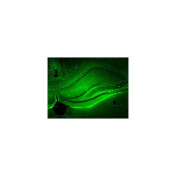
Stereotaxic injection of BrainFectIN/DNA complexes in hippocampus induced an efficient transfection rate of neural cells in all areas of hippocampus.
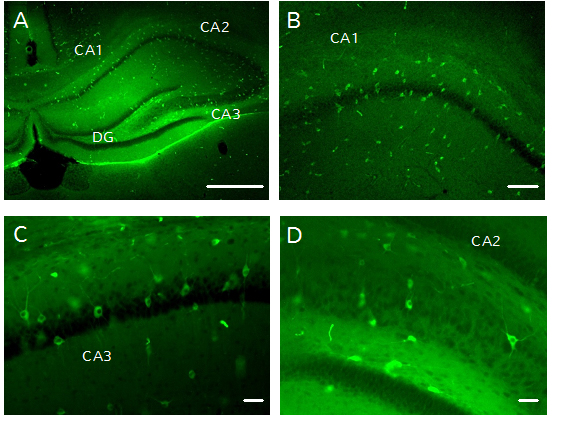
Fig. 1: GFP expression in hippocampus of rat (p11) 48h after BrainFectIN/pGFP injection (ratio 1:1.5). Scale bar = 100µm. The mix was injected through a nanofil needle implanted into hippocampus at the following coordinates: 2mm posterior to the Bregma, 3mm lateral to the midline, and 4mm ventral to the skull surface. Immunochemistry with antibodies directed against GFP was performed in order to increase the signal on PFA fixed brains. GFP+ cells are located in Dentate Gyrus (A) as well as hippocampal areas CA1 (A,B,C), CA2 (A) and CA3 (A,D). Negative control has been done with a stereotaxic injection of DNA alone in the same conditions. It shows a few cells transfected (data not shown).
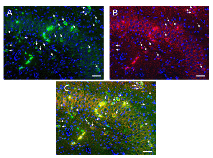
Fig. 2: Double immunofluorescence staining performed in CA3 area. Transfected cells are GFP+ (A, arrows), and interneurons are labelled with GAD 65/67 (B, arrows), nuclei are counterstained with Hoecsht (A,B,C). Merge shows that we are able to transfect GABAergic interneurons (C, arrows). By exclusion, every other cell GFP+ is either pyramidal cell or hippocampal granule cell (C, asterix). It shows that BrainFect allows to transfected at least 3 different neural cell types after intra-hippocampal injection. Scale Bar = 50µm
------------------------------------------------------------------------
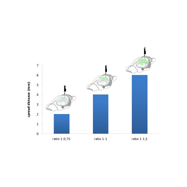
After injection, BrainFectIN™/pGFP complexes can spread into the whole hippocampus structure from rostro-caudal to lateral direction.
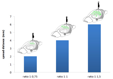
Fig. 3: Quantitative analysis of BrainFectIN™/DNA spread into the rat hippocampus (p12).
---------------------------------------------------------------------------------
GFP expression is detected in ipsi and contra-lateral hippocampus of young rats injected with BrainFectIN™/pGFP complexes 3 weeks after injection.
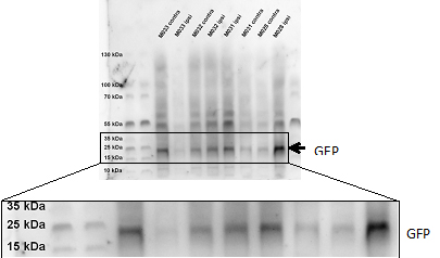
Fig. 4: Qualitative analysis of GFP expression in rat hippocampus (p11) transfected with BrainFectIN™/pGFP. 3 weeks after surgery, hippocampal dissection was performed, ipsi- and contra-lateral hippocampi were pulled apart, proteins were extracted with RIPA buffer, and quantified using Pierce BCA Protein assay kit. Western blot analysis was performed and GFP expression was detected (27 kDa).
Документы
| Название | Код | Цена | ||
| BrainFectIN 100 µL | IV-BF30100 |  |
по запросу | |
| BrainFectIN 250 µL | IV-BF30250 |  |
по запросу | |
| BrainFectIN 500 µL | IV-BF30500 |  |
по запросу |
Информация представлена исключительно в ознакомительных целях и ни при каких условиях не является публичной офертой


